Bladder Scanner
Bladder Scanner代写 The bladder scanner is used to assess and investigate the urine drainage. It is a non-invasive procedurer.

The bladder scanner is used to assess and investigate the urine drainage. Bladder Scanner代写
It is a non-invasive procedure that is used to measure the volumes of urinary bladder. It should be performed as a routine continence assessment or when the retention of urine is suspected. The procedure uses high frequency waves of sound to create images of the inside parts of the body. It uses the ultrasounds than the radiations thus the procedure is safe (Choe, 2007). The instrument is the care standard for the measurement of bladder volume. The instrument is portable, V-mode ultrasonic, battery operated instrument. Other than measuring the volume of the bladder, the bladder scanner also measures the estimated weight of the bladder on instrument basis.
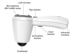
During each of the measurement, the bladder scanner employees the technology of V-mode to obtain several images of aligned B-mode images and create a three-dimension bladder images. It then calculates and display the measurement automatically based upon the generated images thus making it easy to use (Choe, 2007). The measurements of the V-mode tend to be more accurate than the conventional two-dimension ultrasound measurements because they are based on complete multi-faceted images. The photo below describes the parts of the bladder scanner.

The scanhead transmit and receives he ultrasound waves from the body. Bladder Scanner代写
The LCD screen displays the volume of the bladder measurement, setting of gender and the status of the instrument (Moore, 2007). The toggle button toggles the setting of female on and off. When the button is held for five seconds, it displays the number of days up to when calibration is required. In a mobile bladder scan (BVI 6400), holding the top button for three second after performing a measurement of the bladder initiates recording of a voice annotation. The gadget displays the number of days up to when the calibration is required when the Top button is held for five seconds while the bladder scanner is in a charging cradle or docking station. The scan button initiates the imaging of the bladder (Moore, 2007).
The Activation Button activates or resets the bladder scanner Bladder Scanner代写
when needed while the infrared window allows the instrument to communicate with the computer through the docking station. In the BVI 64400 model, the microphone records the voice annotation unto the mobile bladder scanner instrument. It is important to note that before using the bladder scanner for the first time it should have been charged for at least six hours.
Afterwards, Bladder Scanner代写
the instrument can be charged every time the LCD screen shows that the power is low or it is completely drained (Moore, 2007). The LCD displays several icons that have defined meaning. These are best applied by an individual who is well trained to handle the instrument. This is because the instrument should only be used by a person who have been well trained an authorized to handle the machine. The federal laws of the United States restricts the bladder scanner to use by, or on the order of, a physician under the 21 Code of Federal Regulations.
The bladder scanner projects the energy of the ultrasounds through the lower abdomen of the non-pregnant patients to get the bladder images and use them to calculate the volume of the bladder and/or the UEBW non-invasively (Moore, 2007). The instrument is contradicted for fetal use and for use on patients who are pregnant. To ensure the accuracy of the instrument the following conditions should be observed.
The operator of the machine should use care when doing the scan on a patient who have had a pelvic or supra-pubic surgery, surgical incision, scar tissues, sutures and staples since there can be reflection or can affect the transmission of the ultrasound. The operator should not use the instrument on an individual with an open skin or ascites in the supra-pubic region. Also, if the instrument is used on a patient with a catheter in the bladder, the catheter may affect the accuracy of the measurement. Nonetheless, the information obtained from the measurement can still be used for clinical purposes like detection of a blocked catheter (Moore, 2007).
How it Works Bladder Scanner代写
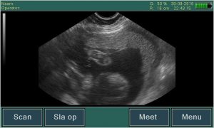
The bladder scanner emits the ultrasounds in one of the plane so that the returning echo appears as a cross section of the organ scanned. The bladder is scanned trans-abdominally using an ultrasound probe on the abdomen (Choe, 2007). This is done at the supra-pubic region. This provides a sagittal and transverse view of the organs scanned. It shows the bladder as a globe-like structure with the interface between the walls of the bladder and the urine indicating a clear segregation. The walls of the bladder have symmetrical, smooth, mildly curved surface. In a transverse scan the shape can vary from almost square to circular.
In women the bladder lies either below or in front of the uterus and a small residual of the urine can be confused with the vagina thus a sagittal view indicates the vagina as a tube-like structure whereas the transverse view shows it to be circular. In men, the pubic bine can slightly abstract the bladder thus it should be viewed at a slightly oblique angle. Following the micturition, the urine residual should not be visible and in adults cannot be identified easily (Alivizatos, 2004).
To do the measurement, Bladder Scanner代写
the instrument should be removed from the cradle and check the battery icon to ensure that it is charged. If the patient is a female then press the Top button to show the female icon. This tells the instrument to filter the uterus from the scan. If the display indicates a female then press the Top button to show a male when doing the scan on a man. One should the clean the scanhead to disinfect it (Alivizatos, 2004).
When the patient is resting in a supine position, palpate the pubic bone and apply the ultrasound on the midline of the abdomen of the patient, 1 inch above pubic bone. The scanhead should then be pressed o the gel pointing downwards towards the expected bladder location. The operator should the press the scan button and hold the bladder scanner steady up to when it beeps once to show the measurement is complete.
Research has shown that non-invasive bladder ultrasound devices like the bladder scanner can assess the volumes of the bladder accurately and reliably (Choe, 2007). Using the bladder scanner avoids many unnecessary catheterizations. Thus using the bladder scanner is considered a preventive measure against infections that are acquired from an indwelling catheter of the urethral. It is also faster and easier to use the ultrasound devices than the catheterization test (Choe, 2007). This method is also favorable to patients with patient with other complications like stroke or spinal cord injury. The results are also easy to interpret and provide valuable results.
References Bladder Scanner代写
Alivizatos, G. e. (2004). Unreliable Residual Volume Measurement After Increased Water Load Diuresis. International Journal of Urology, 1078-81.
Choe, J. e. (2007). Accuracy and Precision of a New Portable Unltrasound Scanner in Residual Urine Volume Measurement. International Urogynecology Journal and Pelvic Dysfunction, 641-644.
Gilbert, R. (2008). bladder Scanner Accuracy During Everyday Use on an Acute rehabilitation. SCI NUrsing, 87-92.
Moore, D. A. (2007). Using a Portable Bladder Scan to Reduce the Incidence of Nosocomial Urinary Tract Infections. Medsurg Nursing, 564-67.


您必须登录才能发表评论。